Health Assessment, part 12: Skin Color Alterations & Edema
Updated: Meris ShuwargerHow to assess alterations in your patient's skin color (e.g., pallor, cyanosis, erythema, jaundice) and how to assess edema (e.g., pitting edema and non-pitting edema)
Full Transcript: Health Assessment, part 12: Skin Color Alterations & Edema
Full Transcript: Health Assessment, part 12: Skin Color Alterations & Edema
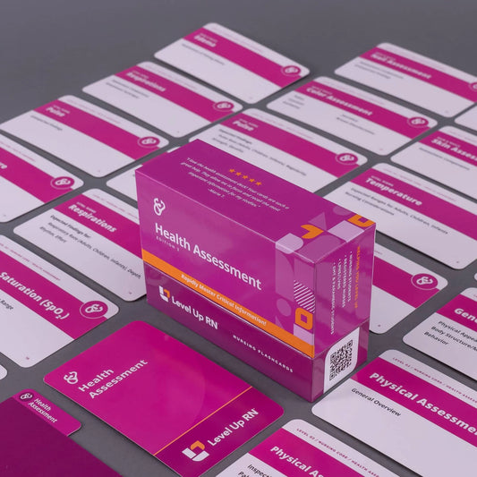
Hi, I'm Meris. And in this video, I'm going to be covering some components of the skin assessment, such as assessing for alterations in skin color, along with assessing for edema. I'm going to be following along using our Health Assessment flashcards. These are available on our website, leveluprn.com. And if you have a set of your own, I would invite you to go ahead and pull them out to follow along with me. All right, let's get started. So first up, we're going to be talking about assessing your patient's skin color for any alterations. And if you look here on the back, we have a nice chart here that shows some different types of alterations and skin color and some potential causes for that. Before we go over this, there's something very important that I want to remind you of, which is that not all patients have white or fair-toned skin. So if I were to present with many of these findings, it would likely be more readily apparent than if there were a patient who had darker skin, brown or black skin, and it is important that you as a healthcare professional are capable and competent to assess for these findings in patients regardless of their skin color. Okay? Really important that you figure out how to identify cyanosis, pallor, jaundice, in your patients who do not have white or fair-toned skin. There are resources freely available online that show different types of presentations of alterations in skin color, in different types of lesions, that sort of a thing. Different sorts of integumentary presentations for certain types of diseases and infections. And I would strongly encourage you to seek out those resources and familiarize yourself with the presentation of these findings in your patients who are not white or fair toned.
All right, that being said, let's get into it. Pallor. Pallor is a fancy way of saying that your patient is pale. And it doesn't mean pale like I am. This is my normal state of being. Pallor refers to that sensation of kind of like-- they don't have any color in their skin. They have been drained of color. So we have on here some causes: anemia, that can do it; shock is a big one - your patients who are in shock very commonly will be pale - and then local arterial insufficiency. It doesn't necessarily have to be a generalized finding. If I have peripheral arterial disease in one limb, it's possible that that limb itself will have pallor. Cyanosis is a blue discoloration of the skin, and this is most commonly associated with hypoxia, hypoxemia due to deoxygenated hemoglobin. Your patients who are blue are close to death, if we do not intervene, typically. So cyanosis is one of those, "Learn it; love it; get a necklace that says it: this kind of a thing means we need to intervene right now." So for instance, if we're talking about the NCLEX testing environment, a patient with cyanosis is going to wildly take priority over a patient with pallor or jaundice. Those things are important to address, but cyanosis means, "Something's wrong with my airway or with my breathing." And that is a concern.
Erythema. If we describe something as, "I noted a patch of erythema on the patient's skin," that means redness. And typically, we are talking about it in a localized place. Typically, I wouldn't say, "That patient was just erythematous head to toe." That's possible, but more likely, I would say, "I saw an erythematous patch on their chest," for instance. I'm saying there's an area of redness. So there's, of course, a lot of causes of it. The biggest is going to be inflammation. And inflammation is often caused by infection as well, so if I see an area that is red, erythematous, I'm concerned, right? I'm going to evaluate further to see if there's a possible source of infection. There's other causes. Generalized erythema could be caused by polycythemia, which is when we have an excess of red blood cells. So that would give me that red or reddy appearance. And then also fever. Anything that makes me hot, I'm going to flush. Alcohol intake for some patients, different kinds of rashes, and then, of course, a sunburn. A burn is going to cause erythema. Jaundice is a yellow discoloration of the skin. And this is most commonly caused by hepatic dysfunction. So your patients with liver disease very commonly may have baseline jaundice or worsening jaundice, or that may be the first sign we see that there's something wrong.
And then brown discoloration. So brown discoloration can be caused by multiple things. If we have sort of a generalized bronzed appearance, that could be caused from Addison's Disease. But if I see brown discoloration in a certain area, that could be what's called stasis dermatitis and can be a sign of venous insufficiency, right? So the blood is pooling in that area. It's not returning to the heart appropriately, and we get that finding called stasis dermatitis, a brown discoloration. Now, when you are assessing for these things, it's very helpful to use mucous membranes, a lot of times. So for instance, talking about patients with darker skin tones, jaundice, I may best see that in the sclera of their eyeballs, right? I may note that yellow discoloration of the eyes. I might see that in my white patients as well, but I may not see that generalized jaundice color. Cyanosis, it's important to look at nail beds; nail beds will turn blue. It's also important to look around the mouth and specifically at mucous membranes. So pull down the lip and assess that mucous membrane. Does it look nice and pink and healthy, or does it look dusky, pale, blue? That's going to be cyanosis. So remember that it's important to not just look from afar, but to really assess your patient for these findings, especially if they do not have white or fair-toned skin.
Okay. Up next, we're going to be talking about edema. And edema is the medical way of saying swelling. Okay? So if something is edematous, that means that it is swollen. So you'll see here-- we have a lot of information on this card. And we say here that it may be characterized as non-pitting or pitting. And what that means is-- non-pitting edema is just kind of that generalized swelling. But if I push down on the skin over a bony prominence and then remove my finger, there's no indentation left. But if I take my finger and I put it over the edematous area - let's say the legs - and I push down over a bony prominence like the tibia. If I do that, I hold down, and then I let go, I'm going to see a pit, a shallow indentation caused by my finger displacing that interstitial fluid. And that's called pitting edema. If our client has non-pitting edema, we're just going to chart it as non-pitting edema. However, if it is pitting, we need some more information, "How severe is that edema? How deep is that pit?" So we've got the little chart here to show you. And the idea here is, again, like I said, we're pushing over a bony prominence. I need something to push back against me. So if I'm trying to assess and I'm pushing here, laterally, it's not going to work. Pushing down over the bone, though, there's that counter pressure so we can really assess for that depth.
Typically, we're looking in the legs. However, you can have edema-- you can have pitting edema anywhere. So it would just be based on the presentation of your patient. Now, we need to document the depth of the indentation. And the way we do this is we-- either 0 or 4+. All the way to 4+, I mean. And what this means is it's telling us how deep it is. 0 means that there is no clinical pitting edema. It doesn't mean that they might not have edema, but it's just not pitting. 1+ means that, after we remove our finger, there's a shallow indentation that is about 2 millimeters in depth. 2+ means it's 4 millimeters in depth; 3+, 6 millimeters; 4+, 8 millimeters or more. And we have a little cool [check-and-hint?] here to help you. And it's just to remember that you should divide the pit depth in millimeters by two to get the pitting level. So it's just by two. And that's to help you remember that. So edema is very common. We see this all the time in patients who just had a baby. We see this in patients with heart failure, patients with fluid volume overload-- there's lots of reasons that we could have a patient with pitting edema. It's important that you document where it is and how severe it is. All right?
Okay. I've got some quiz questions for you to test your knowledge of some key facts I provided in this video: what color would the nurse expect a patient's skin to be, who is experiencing hypoxia? We would expect to see cyanotic skin or blue discoloration. A patient experiencing cirrhosis of the liver presents with yellow discoloration of the skin. How should the nurse document this finding? This would be documented as jaundice. The nurse knows that a patient with 3+ pitting edema has a pit depth of how many millimeters? 6 millimeters. After assessing for edema, the nurse notes a 2-millimeter indentation left in the skin. How should the nurse document this finding? That's 1+ pitting edema. All right, that is it for this video about skin color alterations and assessing for edema. I hope I will see you in the next video where we'll be talking some more about skin assessment. You're doing a really great job, and I'm super proud of you. Thanks for studying with me.


