EKG Interpretation, part 6: Atrial Rhythms

Updated:

In this article, you will learn about the different atrial dysrhythmias, including premature atrial complexes (PACs), atrial flutter, atrial fibrillation and supraventricular tachycardia. The EKG Interpretation video series follows along with our EKG Interpretation Flashcards, which are intended to help RN and PN nursing students study for nursing school exams, including the ATI, HESI, and NCLEX.

Atrial rhythms

The key characteristics of atrial rhythms are abnormal P waves, or P waves that are replaced by flutter or fibrillation waves. Remember from our overview on EKG basics that the expected duration of a P wave is .06-.12 seconds, and the expected amplitude (height) is 2.5mm or 2.5 small boxes.

premature atrial complex on an EKG

Premature atrial complexes (PACs)

Premature atrial complexes (PACs) are a common kind of heart dysrhythmia characterized by premature electrical impulses originating from the atria and occurring too early.

Premature atrial complexes on an EKG

When observing a PAC on an EKG, you will notice that the P wave will appear prematurely. It will have a different look than other P waves, and it may be hidden in the preceding T wave.

Depending on the prematurity of the impulse, PACs can also affect the QRS complex on an EKG. There could be a long pause following the QRS complex, it could cause an abnormally wide QRS complex and can even cause the QRS complex to drop off the EKG completely.

Treatment of PACs

Treatment of PACs is not usually necessary, however, frequent PACs may precede more serious dysrhythmias and they should be monitored closely.

atrial flutter on an EKG

Atrial flutter

Atrial flutter is a type of heart dysrhythmia that occurs when the upper heart chambers (atria) beat faster than the bottom chambers (ventricles).

Atrial flutter on an EKG

When observing atrial flutter on an EKG, the first thing you will notice is that there appear to be multiple sawtooth-like P-waves for every QRS complexes. This means that the atria are beating at a rate 3-4 times the ventricular rate, which is much too fast. Although they may look like P-waves, in atrial flutter these are actually called "flutter waves," or F-waves.

The QRS complexes on an atrial flutter strip will usually be normal and will be under 0.12 seconds in duration.

With atrial flutter, the heart rate will be very, very fast and will be between 250 and 400 beats per minute.

Treatment for atrial flutter

Treatment for atrial flutter can include the use of antiarrhythmic drugs, specifically calcium channel blockers like verapamil and diltiazem. Another treatment option is cardioversion, which is a procedure that restores the heart's normal rhythm by sending electric shocks via electrodes placed on the chest.

Verapamil and diltiazem are two key medications to know for your Pharmacology class, and they are covered in our Pharmacology Flashcards!

atrial fibrillation on an EKG

Atrial fibrillation (Afib)

Atrial fibrillation is a dysrhythmia that can lead to blood clots, stroke, heart failure and more. With atrial fibrillation, the upper chambers of the heart the atria beat very irregularly.

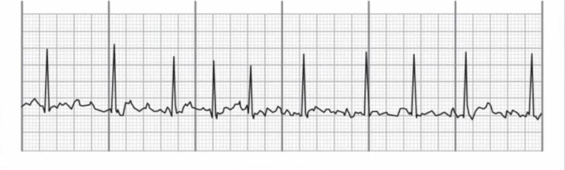
Atrial fibrillation on an EKG

When observing atrial fibrillation on an EKG, the first thing you will notice is that instead of P waves before the QRS complex, there will be lots of little bumps or "fib waves" between QRS complexes.

Distances between R waves will be irregular and the distance between fib waves will vary.

QRS complexes are typically normal in duration at under 0.12 seconds.

The atrial rate for someone with afib will be very fast at between 350 to 600 beats per minute. You will not be able to measure it with your calipers using any box method. You will count the ventricular rate by counting the number of QRS complexes.

Treatment of atrial fibrillation

Patients with afib can be treated with cardioversion as well as antiarrhythmics. Patients with afib are also at high risk for blood clots, stroke and pulmonary embolisms. It is very important that patients receive anticoagulation (blood thinner) medication like warfarin. in order to prevent a blood clot from forming, traveling to the brain, and causing a stroke or thromboembolism.

supraventricular tachycardia on an EKG

Supraventricular tachycardia (SVT)

Supraventricular tachycardia (SVT) describes an abnormally fast heart rate that originates above the ventricles, typically in the atria. "Supra" means above, and ventricular means ventricles, so supraventricular literally means "above the ventricles." Even though ventricular is in the name of this rhythm, it is not a ventricular rhythm, which is a very common mistake.

SVT is considered an umbrella term and includes sinus tachycardia, paroxysmal supraventricular tachycardia, as well as atrial fibrillation and atrial flutter, which we already covered.

Why is sinus tachycardia included in this umbrella if it's a sinus rhythm? Supraventricular tachycardia can actually occur between stretches of normal sinus rhythm.

SVT on an EKG

When observing an SVT on an EKG, the distance between R waves will be equal and pretty regular.

The heart rate for a patient with SVT will be over 100 beats per minute, typically between 150 and 250 beats per minute.

Unfortunately, because the heart rate is so fast, it's hard to evaluate the atrial heart rhythm.

P waves are typically not visible, or if they are, they will be buried in the T wave, making it very difficult to evaluate correctly.

With SVT, the QRS complex will be normal. It will be narrow and under 0.12 seconds in duration.

Treatment for SVT

For treatment of patients with SVT, we can use cardioversion or treat the patient with antiarrhythmics. There is a very special drug to treat SVT, which is not used for any other cardiac arrhythmia: adenosine. This drug is also covered in our Pharmacology Flashcards!

Full Transcript: EKG Interpretation, part 6: Atrial Rhythms

In this video, we are going to talk about atrial rhythms. If you recall, the key characteristic of atrial rhythms is that we have an abnormal P wave or P waves are replaced by flutter or fibrillation waves. So in this video, we're going to talk about premature atrial complexes or PACs. We're going to talk about atrial flutter, atrial fibrillation as well as supraventricular tachycardia. So I'll show you an example with each one, and hopefully, you'll get familiar with how to identify these arrhythmias.

With this strip, we're going to talk about premature atrial complexes or PACs. So you can see that on the right-hand side of the strip, it looks really normal. It's normal sinus rhythm going on over here. But over on this side, this P wave decided to show up early. It didn't wait its turn. It came early, and it's kind of getting all in the T wave's business, the preceding T wave. So that's exactly what a PAC is. It is an abnormal impulse that originates from the atria and occurs early.

So when you are trying to pick out a premature atrial complex, you will notice that the P wave will appear prematurely. It will look different than other P waves, and it may be hidden in the preceding T wave. This is why we have this weird T wave with an extra little lump there.

When you have a PAC, you may end up with a pause following the QRS complex, like we have here. It can also cause the QRS complex to drop completely or cause an abnormally wide QRS complex. So it really depends on the prematurity of the impulse.

So PACs are usually not treated, right? Treatment of PACs is not usually necessary. However, frequent PACs may precede more serious dysrhythmias. So you'll definitely want to keep an eye on them.

Okay, here we have atrial flutter. So you'll notice, right off the bat, that instead of P waves we have these saw-tooth flutter waves instead, or F waves. So it literally looks like a saw. So one way to remember that this is atrial flutter is I think about my husband sawing a log, and I'm checking out his arms, and it makes my heart flutter. So that's my silly, little way of remembering that this is atrial flutter.

When we look at the regularity of this strip, we can see that we have equal distances between these R waves, so the ventricular heart rhythm is regular. Instead of P waves, we have these F waves or flutter waves, which they are also equal distance apart.

If we count the number of small blocks between these F waves, it looks like there is about 5 small blocks. And then if we take 1500 divided by 5, we get 300 beats per minute. So with atrial flutter, the heart rate will be very, very fast. It will be between 250 and 400 beats per minute.

The QRS complexes on an atrial flutter strip will usually be normal, so they will be under 0.12 seconds in duration.

Treatment of atrial flutter can include cardioversion, and we can also use antiarrhythmics to treat this as well. And we will go into more detail about those antiarrhythmics as well as cardioversion at the end of this video series.

Okay, in this strip, we have atrial fibrillation or afib for short. You will notice right off the bat that instead of P waves before the QRS complex, we have all these fib waves or lots of little bumps between the QRS complexes.

So in terms of the rhythm of this strip, if we get our calipers out, we can see that the distance between these R waves is irregular. So we have an irregular ventricular rhythm. And then, instead of P waves, we have these fib waves, the distance between them also varies widely so that is irregular as well.

The atrial heart rate will not be able to be measured by you. It's not like you can get your calipers and figure that out. It will be very fast, so between 350 to 600 beats per minute is common. And then the ventricular heart rate will vary.

You'll notice the QRS complexes are nice and normal so they are under three small boxes in duration or under 0.12 seconds in duration.

In terms of the treatment of AFib, we can treat it with cardioversion as well as antiarrhythmics. And then, the other really important thing that you want to remember is that we want to treat AFib patients with anticoagulation because they are at high risk for getting a thromboembolism and we want to prevent a formation of a clot that can travel to the brain and cause a stroke or a pulmonary embolism, etc. So blood thinners is going to definitely be an important part of this patient's care.

Okay, here we have supraventricular tachycardia or SVT. So SVT really describes any abnormally fast heart rate that originates above the ventricles, typically in the atria which is why I included it here when we're talking about atrial rhythms. So under this SVT umbrella. This includes sinus tachycardia, atrial fibrillation, as well as atrial flutter.

So when we look at this rhythm, it is actually pretty regular in terms of the ventricular heart rhythm. So if you measure between these R waves, it will be equal distance apart.

We can't really evaluate the atrial heart rhythm because the P waves are not really visible. You can see here that this T wave looks a little funky because this P wave is kind of buried into the side of it because this heart rate is so, so fast.

We can't really evaluate the P wave because it's not typically visible when we're talking about SVT. And therefore, we also can't evaluate the atrial heart rhythm.

So the heart rate with SVT will be over 100 beats per minute. It is typically between 150 and 250 beats per minute. With SVT, the QRS complex will be normal. It will be nice and narrow, under three small boxes or 0.12 seconds.

In terms of treatment of SVTs, we can use cardioversion or treat the patient with antiarrhythmics.

Back to blog

Leave a comment

Please note, comments need to be approved before they are published.